Tuesday, April 12, 2022

DENTAL ANESTHESIA

 Around 10 to 30 percent

. Trusted Source of people have anxiety and concerns about pain with dental procedures. Anxiety can delay getting treatment and that can make the problem worse.

Anesthetics have been around for over 175 years! In fact, the first recorded procedure with an anesthetic was done in 1846 using ether. 

We’ve come a long way since then, and anesthetics are an important tool in helping patients feel comfortable during dental procedures.With lots of different options available, anesthesia can be confusing. We break it down so you’ll feel more confident before your next dental appointment.

The Different Types of Dental Anesthesia

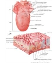
Different types of dental sedations can be used for dental procedures and surgeries. Still, many people are apprehensive about being unconscious during a procedure.  If this sounds like you, you can remain conscious during a procedure and not feel pain and anxiety.

Local Anesthesia

Local anesthesia used in dental clinics such as Novocaine is injected into the gum line. The area will feel completely numb in as little as a few minutes. You will remain awake and sedated, but you will feel reduced sensations and no pain throughout the procedure. Since dental procedures are mostly not time-consuming and are outpatient procedures, local anesthesia is commonly used. However, lengthier procedures may require the patient to be sedated for longer.

Nitrous Oxide

The dentist combines a local anesthetic with nitrous oxide or laughing gas. You breathe in the medication and gas through a mask. The mask stays on for the duration of the procedure. You will remain conscious throughout the procedure, albeit sedated.

Oral Sedation 

The dentist provides medication to take orally before the procedure. we have many dosage can be achieved. Minimal sedation will put you in a dreamlike state, however, still very much awake. Moderate and deep sedation may make you fall 
asleep, but it should be easy to wake you.

IV Sedation

 Another delivery method of a sedative is intravenously or through a vein. With IV sedation, you will be in a semi-cinscious state and have little or no memory of the actual surgery.

What are the side effects of dental anesthesia?

Side effects of dental anesthesia depend on the type of anesthetic used. General anethesia risker than local anesthesia or sedation.

  • nausea or vomiting

    • headache

      • sweating or shivering

        • hallucinations, delirium, or confusion

          • slurred speech

            • dry mouth or sore throat

              • pain at the site of injection

                • dizziness

                  • tiredness

                    • numbness

                    • lockjaw (trismus) caused by trauma from surgery; the jaw opening is temporarily reduced
                      Vasoconstrictors such as epinephrine added to anesthetics can also cause heart and blood pressure problems.
                      These are some reported side effects of anesthetics. Ask your dental care team about your specific medication and any concerns you may have about the medication.


                      Saturday, March 19, 2022

                      DENTAL VENEERS


                      What are Dental Veneers?

                      Dental veneers or porcelain veneers or dental porcelain laminates are wafer-thin, custom-made shells of tooth-colored materials designed to cover the front surface of teeth to improve your appearance.

                       These shells are bonded to the front of the teeth changing their color, shape, size, or length.

                      it;s a possible solution to help you achieve the look you desire. They are a popular choice for those with chipped teeth, a gap between teeth, or misshaped teeth.


                      Veneers can be used to correct a wide range of dental issues, such as:

                      • teeth that are stained and can’t be whitened by bleaching
                      • chipped or worn teeth
                      • crooked or misshapen teeth
                      • uneven spaces or a large gap between the upper front teeth


                      Types of Dental Veneers

                      Dental veneers can be made from porcelain or from resin composite materials. Porcelain veneers resist stains better than resin veneers. They also better mimic the light-reflecting properties of natural teeth. You will need to discuss the best choice of veneer material for you with your dentist.

                      Preparation

                      To prepare a tooth for a veneer, your dentist will reshape the tooth surface, which is an amount nearly equal to the thickness of the veneer to be added to the tooth surface. You and your dentist will decide whether they numb the area before trimming off the enamel. Next, your dentist will make a model, or impression, of your tooth. This model is sent out to a dental laboratory, which makes your veneer. It usually takes 2-4 weeks for the veneers to come back from the laboratory. Temporary dental veneers can be used in the meantime.

                      < dir="ltr" style="box-sizing: inherit; letter-spacing: -0.273104px; margin: 0px 0px 0.715em;">

                      Bonding

                      Your dentist will place the veneer on your tooth to examine its fit and color, repeatedly removing and trimming the veneer to achieve the proper fit, before permanently cementing it to your tooth. The veneer color can be adjusted with the shade of cement to be used. Next, to prepare your tooth to receive the veneer, your tooth will be cleaned, polished, and etched. Etching roughens the tooth to allow for a strong bonding process. A special cement is applied to the veneer and it is then placed on your tooth. Once the veneer is properly positioned, your dentist will shine a special light beam on it to activate chemicals in the cement, causing it to harden very quickly. The final steps involve removing any excess cement, checking your bite and making any needed adjustments. Your dentist may ask you to return for a follow-up visit in a couple of weeks to check your gums and the veneer's placement.

                      Dental Veneer Benefits

                      Veneers offer these advantages:

                      • They provide a natural tooth appearance.

                      • Gums tolerates porcelain well.

                      • Porcelain veneers are stain resistant.

                      • A color can be selected to make dark teeth appear whiter.

                      • They generally don't require as much shaping as crowns do, yet they are stronger and look better.

                      Dental Veneer Risks

                      Downsides to dental veneers include:

                      • The process cannot be undone.

                      • Veneers cost more than composite resin bonding.

                      • Veneers usually cannot be repaired if they chip or crack.

                      • Because enamel has been removed, your tooth may become more sensitive to hot and cold foods and drinks.

                      • Veneers may not exactly match the color of your other teeth. Also, the veneer's color cannot be altered once it’s in place. If you plan on whitening your teeth, you need to do so before getting veneers.

                      • Though not likely, veneers can dislodge and fall off. To minimize the chance of this occurring, do not bite your nails, chew on pencils, ice or other hard objects, or otherwise put too much pressure on your teeth.

                      • Teeth with veneers can still experience decay, possibly necessitating full coverage of the tooth with a crown.

                      • Veneers are not a good choice for people with unhealthy teeth (for example, those with decay or active gum disease), weakened teeth (as a result of decay, fracture, large dental fillings), or for those who don't have enough existing enamel on the tooth surface.

                      • People who clench and grind their teeth are poor candidates for porcelain veneers, as this can cause the veneers to crack or chip.

                       Before you get veneers

                      • Your teeth and gums must be healthy before you get veneers. Your dentist can treat any disease or decay before your veneers are placed.
                      • Veneers are not always a good choice for patients who clench or grind their teeth, because the thin veneers may chip or break. If you clench or grind your teeth, your dentist may suggest you wear a plastic dental night guard while sleeping.
                      • Although your dentist removes as little tooth enamel as possible for veneers, the process cannot be undone once the enamel is removed.
                      • It is possible for veneers to come loose over time. In that case, new ones might be needed.
                      • As with all your dental care, discuss all your expectations and treatment options with your dentist. Regular dental visits are a must for keeping your teeth and gums healthy.

                      After you get veneers

                      • Veneers can chip or break under pressure. Avoid biting your fingernails and chewing on hard objects, such as pencils or ice.
                      • It may take you a few days to get used to the feel of your veneers. But do let your dentist know if your bite does not feel right after the veneer has been placed. He or she will correct it before you leave the office.
                      • Keep your teeth and gums clean by brushing and flossing each day. You can still get cavities under or around veneers. Look for dental care products that show the American Dental Association’s Seal of Acceptance. This says that a product has met ADA standards for safety and effectiveness.

                      Friday, March 18, 2022

                      TEETH WHITENING

                       How do I get a bright smile?

                      Brushing, mouth wash and flossing are everyday ways to keep your teeth bright, white and healthy. If you might feel like your smile is lacking sparkle or is more yellow than it used to be, you're not alone. When the American Academy of Cosmetic Dentistry asked people what they'd most like to improve about their smile. The American Association of Orthodontists also found the nearly 90% of patients requested tooth whitening.

                      Thinking about teeth whitening? Get the facts first. Here are five of the most commonly asked questions about the process.


                      Why Did Teeth Change Color?

                      we have number of reasons:

                      Food and Drink
                      Coffee, tea are some major staining culprits. What do they have in common? Intense color pigments called chromogens that attach to the white, outer part of your tooth (enamel).

                      Tobacco Use
                      Two chemicals found in tobacco create stubborn stains: Tar and nicotine. Tar is naturally dark. Nicotine is colorless until it’s mixed with oxygen. Then, it turns into a yellowish, surface-staining substance.

                      Age
                      Below the hard, white outer shell of your teeth (enamel) is a softer area called dentin. Over time, the outer enamel layer gets thinner with brushing and more of the yellowish dentin shows through.

                      Trauma
                      If you’ve been hit in the mouth, your tooth may change color because it reacts to an injury by laying down more dentin, which is a darker layer under the enamel.

                      Medications
                      Tooth darkening can be a side effect of certain antihistamines, antipsychotics and high blood pressure medications. Young children who are exposed to antibiotics like tetracycline and doxycycline when their teeth are forming, may have discoloration of their adult teeth later in life. Chemotherapy and head and neck radiation can also darken teeth.

                       How do teeth whitening products work?

                      There are a ton of different professional teeth whitening products out there, but they all use the same basic ingredients; peroxides. Peroxides, such as hydrogen peroxide and carbamide peroxide, contain very volatile oxygen molecules.

                      When these oxygen molecules touch surface stains on your teeth, they react by breaking apart the chemical bonds that hold the stains onto your enamel. This “bleaches” them away and restores the appearance of your smile.

                      Every professional teeth whitening product uses hydrogen peroxide or carbamide peroxide as the active whitening ingredient. However, other ingredients are often used alongside peroxides, such as fluoride. Adding fluoride to a teeth whitener helps prevent the weakening of your teeth during the process, and also minimizes the risk of tooth sensitivity after teeth whitening. 


                      Does Whitening Work on All Teeth?

                      No, which is why it’s important to talk to your dentist before deciding to whiten your teeth, as whiteners may not correct all types of discoloration. For example, yellow teeth will probably bleach well, brown teeth may not respond as well and teeth with gray tones may not bleach at all. Whitening will not work on caps, veneerscrowns or fillings. It also won’t be effective if your tooth discoloration is caused by medications or a tooth injury.

                      Are There Any Side Effects from Teeth Whitening?

                      Some people who use teeth whiteners may experience tooth sensitivity. That happens when the peroxide in the whitener gets through the enamel to the soft layer of dentin and irritates the nerve of your tooth. In most cases the sensitivity is temporary. 

                      Overuse of whiteners can also damage the tooth enamel or gums, so be sure to follow directions and talk to your dentist.


                      Thursday, March 17, 2022

                      TONGUE

                      •  Tongue: a musclar structure in the oral cavity, divided in to 2 parts:

                      1.  Oral (pesulcal), movable part-the anterior 2\3 of the tongue 
                      2. Pharyngeal (postsulcal), nonmovable part-the posterior 1\3 of the tongue

                      • The sulcus terminalis, a V-shaped groove immediately posterior to the circumvallate papilla, separates the oral part from the pharyngeal part of the tongue
                      • The foramen cecum, which was the initial development site for the thyroid gland, is located at the tip (or angle) of the V.
                      • Median fibrous septum separates the tongue into halves 

                       FUNCTIONS

                      • Mastication 
                      •  Taste
                      •  Talking
                      • Degultition

                      APPEARANE 

                      • The tongue typically is pink and and covered with numerous small bumps called papilla
                      • Change in color or texture may reflect health problems:

                      1. Leukoplakia
                      2. Squamous cell carcinoma
                      3. Nutritional deficiencies

                      • An unusual appearance of the tongue may represent a benign harmless condition:

                      1. Fissured toungue
                      2. Bkack hairy tongue
                      3. Geographic toungue

                      MUSCLE TYPES

                      • Extrinsic-move the tongue as an anatomic structure 
                      • Intrinsic-change the tongue's shape   


                      geographic tongue


                      hairy tongue 
                      fissured tongue


                      ORAL PART (PRESULCAL)

                      ·         The oral part of the tongue is also known as the anterior 2\3 of the tongue

                      ·         Develops from the 2 lateral lingual swelling and tuberculum impar of the 1st pharyngeal arch-thus mucosa is innervated by the nerve of the 1 st arch, which is the trigeminal nerve (lingual n.)

                      ·         Has a dorsal and a ventral surface

                      Dorsal Surface

                      ·         The mucosa is keratinized stratified squamous epithelium

                      ·         Is bounded posteriorly by the sulcus terminalis

                      Structures

                      Comments

                      Median sulcus

                      A groove that travels anteriorly in the midline

                      Represents the location of the median septum that divides the tongue in halves

                      The septum is thicker posteriorly but thinner anteriorly

                      Papillae-there are 4 types of papillae on the dorsal surface of the tongue:

                      ·         Filiform-lack tast buds

                      Most numerous type of papillae

                      Have thick keratin on epithelium

                      ·         Fungiform-have taste buds that receive taste innervation from the facial nerve (chorda tympani branch)

                      Scattered throughout the dorsum of the oral part

                      Lack keratin on the epithelium

                      ·         Foliate-have taste buds that receive taste innervation from the facial nerve (chorda tympani branch)

                      Are located on the side of the tongue in 4 to 5 folds immediately anterior to the palatoglossal fold

                      Lack keratin on the epithelium

                      ·         Circumvallate-have taste buds that receive tast innervation from the glossopharyngeal nerve

                      Generally a nonkeratinized epithelium

                      Lie in a row immediately anterior to the sulcus terminalis

                      Papillae are raised projections that increase the surface area

                      5 basic types of taste are differentiated by the taste buds:

                      ·         Bitter

                      ·         Salt

                      ·         Sweet

                      ·         Sour

                      ·         Umami

                      Glands

                      There are numerous mucous and serous glands on the dorsal surface


                      Ventral Surface

                      ·         The mucosa is nonkeratinied stratified squamous epithelium

                      Structures

                      Comments

                      Lingual frenulum

                      A midline fold of tissue

                      Connect the ventral surface of the tongue to the floor of the oral cavity

                      Sublingual papilla

                      A swelling on both sides of the lingual frenulum at the tongue base

                      Marks the entrance of saliva from the submandibular glands into the oral cavity

                      Continuous with the sublingual folds overlying the sublingual glands on the floor of the oral cavity

                      Plica fimbriata

                      Fimbriated folds

                      Lateral to the lingual frenulum

                      Deep lingual veins

                      Can be observed through the mucosa between the plica fimbriata and the lingual frenulum


                      PHARYNGEAL PART(POSTSULCAL)

                      ·           The pharyngeal part of the tongue is also known as the posterior 1\3 of the tongue

                      ·         Develops from the hypobranchial eminence of the 3rd pharyngeal arch-thus mucosa is innervated by the nerve of the 3rd arch (the glossopharyngeal nerve)

                      ·         The area immediately posterior to the palatoglossal folds (alsa called the anterior pillar of the fauces) is the oropharynx

                      ·         Has a dorsal surface only

                      ·         Does not possess any papilla

                      Dorsal Surface

                      ·         The mucosa is nonkeratinized stratified squamous epitheelium

                      Structures

                      Comments

                      Lingual tonsils

                      Large nodules of lymphatic tissue

                      Cover the pharyngeal surface of the tongue

                      Glosseoepiglottic folds

                      Mucous membrane of nonkeratinized stratified squamous epithelium from the pharyngeal  part and lateral wall of pharynx that reflects onto the anterior epiglottis, forming:

                      ·         Median glossoepiglottic fold

                      ·         2 lateral glossoepiglorric folds

                      The median glossoepiglottic fold is bordered by a depression on each side:

                      ·         Vallecula

                      Connect the posterior portion of the pharyngeal part of the tongue with the epiglottis of the larynx


                      Muscle

                      Origin

                      Insertion

                      Action

                      Nerve

                      Comment

                      Genioglossus

                      Superior genial tubercle of the mandible

                      Superior fibers fan into the entire ventral surface of the tongue while intermixing with the (intrinsic muscles)

                      Intermediate fibers fan posteriorly in attach to the posterior tongue

                      Inferior fibers insert into the body of the hyoid via an aponeurosis

                      Protrusion of the tongue

                      Bilaterally-the 2 muscles will depress the central

                      Portion of the tongue, which makes the dorsal surface concave

                      Unilaterally-makes the tongue deviate to the contralateral side

                      Hypoglossal n.

                      The lingual a. is located between the genioglossus and hyoglossus mm.

                      Hyoglossus

                      Greater cornu and anterior portion of the body of the hyoid

                      Lateral portion of the tongue between the styloglossus m. and the interior longitudinal m.

                      Depresses the tongue


                      The lingual n., hypoglossal n., and submandibular duct are located on the lateral surface of the hyoglossus m.

                      Some authors describe the chondroglossus asa separate muscle or as part of the hyoglossus

                      Styloglossus

                      Anterolateral portion near the apex of the styloid process

                      Stylomandibular ligament

                      Longitudinal portion inserts into the dorsolateral part of the tongue to intermix with the inferior longitudinal m.

                      Oblique portion inserts into the dorsolateral portion of the tongue to intermix with the hyoglossus m.

                      Retrusion of the tongue

                      Elevation of the tongue


                      Smallest of the extrinsic tongue muscles

                      Palatoglossus

                      Palatine aponeurosis (oral surface)

                      Lateral side of the tongue where some fibers intermix with the transverse m. and some along the dorsal surface of the tongue

                      Elevation of the root of the tongue

                      Narrows the oropharyngeal isthmus for deglutition

                      Pharyngeal plexus (the motor portion of this plexus is formed by the pharyngeal branch of the vagus n.)

                      Grouped as either an extrinsic tongue muscle of the tongue or a muscle of the soft palate


                      Muscle

                      Origin

                      Insertion

                      Actions

                      Nerve

                      Comment

                      Superior longitudinal

                      Median septum

                      Submucous layer near epiglottis

                      Lingual margins

                      Shortens

                      Curls the tongue’s apex and lateral margins upward, which makes the dorsal surface concae

                      Hypoglossal n.

                      Located immediately deep to the mucous membrane of the tongue’s dorsal surface

                      Inferior longitudinal

                      Root of the tongue

                      Body of the hyoid

                      Apex of the tongue

                      Shortens

                      Curls the tongue’s apex downward, which makes the dorsal surface convex

                      Runs the length of the tongue between the hyoglossus and genioglossus mm.

                      Transverse

                      Median septum

                      Fibrous tissue in the submucosa of the lingual margins

                      Some fibers intermix with palatoglossus

                      Narrows lengthens

                      Runs the width of the tongue

                      Vertical

                      Submucosa of dorsal surface of tongue

                      Submucosa of ventral surface of tongue

                      BROADENS

                      Flattens

                      Runs from the dorsal to the ventral tongue surface


                      TYPES OF SENSORY NERVE SUPPLY

                      Type

                      Function

                      Nerves

                      General somatic afferent (GSA)

                      Pain, temperature, discriminative touch

                      Trigeminal (via lingual), glossopharyngeal, and vagus (via internal laryngeal), to innervate the mucosa

                      Special visceral afferent (SVA)

                      Taste

                      Facial (via chorda tympani), glossopharyngeal, and vagus (via internal laryngeal), to innervate the tast buds


                      GENERAL SENSORY INNERVATION (GENERAL SOMATIC AFFERENT)

                      Nerve

                      Source

                      Course

                      Lingual

                      Mandibular division of the trigeminal n.

                      Lies inferior to the lateral pterygoid m. amd medial and anterior to the inferior alveolar nn. Within the infratemporal fossa

                      Chorda tympani branch of the facial n. joins its posterior part

                      Lingual n. passes between the medial pterygoid m. and the ramus of the mandible to pass obliquely, entering the oral cavity bounded by the superior pharyngeal constrictor m., the medial pterygoid, and the mandible

                      Enters the oral cavity lying along the lingual tuberosity of the mandible

                      The submandibular ganglion is supended from the lingual n. at the posterior border of the hyoglossus m.

                      Continues anteriorly and passes onto the lateral surface of the hypoglossus

                      Passes from the lateral side inferiorly and medial to the submandibular duct to reach the mucosa of the tongue

                      Supplies GSA fibers to the epithelium and papillae of the tongue’s anterior 2\3, mucosa along the floor of the oral cavity (linguoalveolar ridge), and gingiva on the lingual aspect of the mandibular teeth

                      Glossopharyngeal

                      Arises as a cranial nerve from the medulla oblongata

                      Passes through the jugular foramen with the vagus and accessory nn.

                      Within the foramen, it passes between the internal carotid a. and the internal jugular v.

                      Continues inferiorly and posteriorly relative to the stylopharyngeus m.

                      Passes anteriorly with the stylopharyngeus m. and travels between the superior and middle constrictor mm. to beome located by the palatine tonsils

                      Small lingual branches distribute GSA fibers to the epithelium of the tongue’s posterior 1\3, in addition to the fauces

                      Internal laryngeal

                      Superior laryngeal branch of the vagus n.

                      Vagus n. branches from the medulla oblongata and passes through the jugular for amen with the glossopharyngeal and accessory nn.

                      Within the foramen, it passes between the internal carotid a. and the internal jugular v.

                      A series of nerves branch from the vagus in the neck, including the superior laryngeal n., which travels inferiorly posterior to the internal carotid a. on the side of the pharynx and divides into the internal and external laryngeal nn.

                      The internal laryngeal n. passes inferior to the larynx and passes through the thyrohyoid membrane with the superior laryngeal vv.

                      Distributes GSA fibers to the tongue’s base at the epiglottic region and the mucous membranes of the larynx as far inferiorly as the false vocal folds


                      SPECIAL SENSORY INNERVATION (SPECIAL VISCERAL AFFERENT)

                      Nerve

                      Source

                      Course

                      Chorda tympani

                      Facial n. in the tympanic cavity

                      Carries preganglionic parasympathetic fibers to the submandibular ganglion and tast fibers to the anterior 2\3 of the tongue

                      Passes anteriorly to enter the tympanic cavity and lies along the tympanic membrane and malleus until exiting the petrotympanic fissure

                      Joins the posterior border of the lingual n.

                      Lingual n. is distributed to the anterior 2\3 of the tongue, and the SVA fibers from the chorda tympani travel to the taste buds in this region

                      Glossopharyngeal

                      Arises as a cranial nerve from the medulla oblongata

                      Passes through the jugular foramen with the vagus and accessory nn.

                      Within the foramen, it passes between the internal carotid a. and the internal jugular v.

                      Continues inferiorly and travels posterior to the stylopharyngeus m.

                      Passes anteriorly with the stylopharyngeus m. and travels between the superior and middle constrictor mm., to be located by the palatine tonsils

                      Small lingual branches distribute SVA fibers to the taste buds in the mucous membrane of the tongue’s posterior 1\3 and the circumvallate papilla

                      Internal laryngeal

                      Superior laryngeal branch of the vagus n.

                      Vagus n. branches from the medulla oblongata and passes through the jugular foramen with the glossopharyngeal and accessory nn.

                      Within the foramen, it passes between the internal carotid a. and the internal jugular v.

                      A series of nerves branch from the vagus in the neck, including the superior laryngeal n., which travels inferiorly posterior to the internal carotid on the side of the pharynx and divides into the internal and external laryngeal nn.

                      The internal laryngeal n. passes inferior to the larynx and passes through the thyrohyoid membrane with the superior laryngeal vv.

                      Distrbutes SVA fibers to the taste buds scattered at the base of the tongue at the epiglottic region


                      Nerve

                      Sorce

                      Course

                      Hypoglossal

                      Arises as a series of rootlets from the medulla oblongata and passes through the hypoglossal canal

                      Travels inferiorly and is located between the internal carotid a. and the internal jugular v.

                      Passes anteriorly as it wraps around the occipital a.

                      Passes superficial to the external carotid and the loop pf the lingual a. in its anterior path

                      Passes deep to the posterior belly of the digastric and the stylohyoid m. and lies superficial to the hyoglossus m. with the accompanying vein of the hypoglossal n.

                      Passes deep to mylohyoid m. and continues anteriory in the genioglossus m.

                      Muscular branches supply:

                      ·         All intrinsic tongue muscles

                      ·         Hyoglossus m.

                      ·         Styloglossus m.

                      ·         Genioglossus m.

                      Pharyngeal  plexus

                      Pharyngeal plexus (the motor portion of this plexus is formed by the pharyngeal branch of the vagus n.)

                      Arises from the upper part of the inferior ganglion of the vagus n.

                      Lies along the upper border of the middle constrictor m., where it forms the pharyngeal plexus

                      Motor branches from the plexus are distributed to the muscles of the pharynx and soft palate (with the exception of the tensor veli palatine m.)

                      It the tongue, it innervates:

                      ·         Palatoglossus m


                      Artery

                      Source

                      Course

                      Lingual

                      External carotid a. within the carotid triangle

                      Passes superiorly and medially (obliquely) toward the greater cornu of the hyoid bone and makes a loop by passing anteriorly and inferiorly while traveling superficial to the middle constrictor m.

                      While forming a loop, the artery is crossed superficially by the hypoglossal n.

                      Passes deep to the posterior belly of the digastric m. and the stylohyoid m. as it travels anteriorly, where it gives off a suprahyoid branch that travels on the superior surface of the hyoid bone, supplying the muscles in that area

                      The lingual a. passes deep to the hyoglossus m. and travels anteriorly betweem it and the genioglossus m.

                      After passing deep to the hyoglossus, 2 to 3 small dorsal lingual aa. Are given off at the posterior border of the hyoglossus.

                      The lingual a. continues to pass anteriorly and gives off the sublingual branch at the anterior border of the hyoglossus

                      The deep lingual a., the terminal branch or continuation of the lingual a. once the sublingual a. is given off, travels superiorly to reach the tongue’s vental surface

                      Dorsal lingual

                      Lingual

                      After passing deep to the hyoglossus, 2 to 3 small dorsal lingual aa. Are given off at the posterior border of the hyoglossus; they pass in a superior direction to the posterior 1\3 of the dorsum of the tongue and provide vascular supply to the mucous membrane in this region, the palatoglossal arch, the palatine tonsil, the epiglottis, and the surrounding soft palate

                      Deep lingual

                      The deep lingual a., the terminal branch or continuation of the lingual a. once the sublingual a. is given off, travels superiorly to reach the tongue’s ventral surface

                      Located between the inferior longitudinal m. of the tongue and the mucous membrane, the deep lingual a. is accompanied by branches of the lingual n., and it anastomoses with its counterpart from the other side

                      Sublingual

                      The sublingual branch arises at the anterior border of the hyoglossus

                      The sublingual a. passes anteriorly between the genioglossus and mylohyoid mm. to the sublingual gland and provides vascular supply to the gland and the muscles and mucosa in the area

                      Typically has 2 branches of significance:

                      ·         Branch that passes mylohyoid to anastomose with submental a.

                      ·         Branch that passes deep to the gingiva to anastomose with branch from contralateral side

                      ·         At this anastomosis, typically 1 branch (although may be multiple branches) arises to enter a small lingual foramen superior to the genial tubercles in the posterior midline

                      Submental

                      A branch of the facial a. from the external carotid a.

                      Given off at the submandibular gland, travels anteriorly superficial to the mylohyoid m.

                      Anastomoses with a branch from the sublingual branch of the lingual a. to help supply the tongue

                      Tonsillar

                      While ascending superiorly along the lateral side of the pharynx, it passes into and supplies the superior constrictor m. until reaching the palatine tonsil and root of the tongue

                      Ascending pharyngeal

                      External carotid a.

                      The smallest branch arising from the external carotid a.

                      Ascends superiorly between the lateral aspect of the pharynx and the internal carotid a.

                      Has branches supplying the palatine tonsil that anastomose with tonsillar branch of facial and dorsal lingual branches at root of the tongue


                      Vein

                      Course

                      Lingual

                      Receives tributaries from the deep lingual vv. On the ventral surface, and the dorsal lingual vv. From the dorsal surface

                      Passes with the lingual a., deep to the hyoglossus m., and ends in the internal jugular v.

                      The vena comitans nervi hypoglossi, or accompanying vein of the hypoglossal n., begins at the tongue’s apex and may either join the lingual v. or accompany the hypoglossal n. and enter the commen facial v., which empties into the internal jugular

                      Submental

                      Anastomoses with the branches of the lingual v.

                      Parallels the submental a. on the superficial surface of the mylohyoid m. and ends in the facial v.


                      ANKYLOGLOSSIA

                      • Ankyloglossia: condition in which the lingual frenulum is restricted because of an increase in tissue, leading to reduced tongue mobility
                      • Also known as tongue-tie
                      PRESENTATIONS
                      • Tongue may not be capable of protrusion beyond the incisors
                      • Tongue may not be capable of touching the palate 
                      • Tongue may have a V-shaped notch at its tip or may appear bilobed on protrusion
                      COMPLICATIONS
                      • Causes problems for babies who breastfeed
                      • If the tongue cannot clear the oral cavity of food, caries, periodontal disease, and halitosis can rresult
                      • If condition is severe, can cause a speech impediment
                      TREATMENT
                      • If necessary, the lingual frenulum may be cut (frenectomy)

                      HYPOGLOSSAL NERVE  PARALYSIS 

                      • Hypoglossal nerve lesions paralyze the tongue on 1 side 
                      • On protrusion, the tongue deviates to the ipsilateral (same) or contralateral side, depending on the lesion site
                      LOWER MOTOR NEURON LESION
                      • Lesions to the hypoglossal nerve cause paralysis on the ipsilateral side:
                      1. Tongue deviates to the paralyzed side on protrusion (the paralyzed muscles will lag, causing the tip to deviate)
                      2. Musculature atrophies on the paralyzed side 
                      3. Tongue fasciculations occure on the paralyzed side 
                      Example: With a neck wound that cuts the right hypoglossal nerve, the tongue deviates to the right on protrusion, and the right half of the tongue will later demonstrate atrophy and fasciculations

                      UPPER MOTOR NEURON LESION

                      • Causes paralysis on the contralateral side:
                      1. Tongue deviates to the side opposite the lesion
                      2.  Musculature atrophies on side opposite the lesion
                      Example: After a stroke on the right side of the brain that affects the right upper motor neurons, the tongue deviates to the left on protrusion, and the left half of the tongue will atrophy


                      SQUAMOUS CELL CARCINOMA

                      • Squamous cell carcinoma accounts for most cancers of the oral cavity 
                      • In the tonge, usually located on the anterolateral aspect
                      • Alcohol and tobacco use are risk factors
                      • Premalignant lesion, such as erythroplasia and leukoplakia, should be identified, because early diagnosis and treatment are paramount for long-term survival
                      • Radiographic imaging helps reveal the tumor's extent and location 
                      • Staging of the tumor guides prognosis
                      TREATMENT

                      • Excision or radiation therapy, or possibly in combination with chemotherapy
                      • If lesion is detected early, excision may suffice 
                      • With later tumor stages, a 2nd primary squamous cell carcinoma must be excluded

                      LEUKOPLAKIA

                      • Leukoplakia: a commen premalignant condition of the oral cavity involving the formation of white spots on the mucous membranes of the tongue and inside the mouth 
                      • Hairy leukoplakia is a type observed in persons with compromised immune systems
                      • Risk factors:
                      1. Tobacco use
                      2. Alcohol use
                      3. Human immunodeficiency virus (HIV) infection
                      4. Epstein-Barr virus infection
                      • Although a precancerous lesion, it may not progress to oral cancer

                      Pericoronitis

                      Defination of pericoronitis:   Pericoronitis is swelling and infection of the gum tissue around the lower wisdom teeth, lower wisdom teeth t...